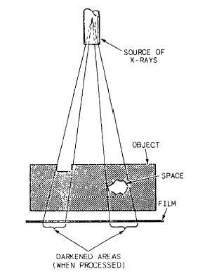
Figure 15-10.—Diagram of radiographic exposure.
and oriented with respect to penetrating rays to be
reliably detected. Radiography is one of the most
expensive and least sensitive methods for crack
detection. It should only be used to detect flaws that are
not accessible or favorabl y oriented for use by other test
methods.
The extent of recorded information
upon the following three prime factors:
1. The composition of the material.
is dependent
2. The product of the density and the thickness of
the material.
3. The energy of the X-rays, which is incident upon
the material. Material discontinuities cause an apparent
change in these characteristics, and thus make
themselves detectable.
Figure 15-10 is a diagram of radiographic exposure
showing the elements of the system. Radiation passes
through the object and produces an invisible or latent
image in the film. When processed, the film becomes a
radiograph or shadow picture of the object. Since more
radiation passes through the object where the section is
thin or where there is a space or void, the corresponding
area on the film is darker. The radiograph is read or
interpreted by comparing it with the known nature of the
object.
RADIOGRAPHIC INTERPRETATION.—The
usefulness of the information obtained from the
radiographic process depends upon the intelligent
interpretation of the derived image. To successfully
interpret the radiograph, the radiographic interpreter
must have a working knowledge of the component or
material and be able to relate the images to the
conditions likely to occur. Specifications are used to
spell out the discontinuities that maybe considered
detrimental to the function of the part and the acceptable
magnitudes of the discontinuities. It is the duty of the
film interpreter to recognize the various discontinuities,
their magnitudes, and be capable of relating them to the
particular specification required. The responsibility and
capability of the radiographic interpreter cannot be
overemphasized. Often, many human lives and
investments of millions of dollars are depending on the
judgement of the radiographic interpreter.
RADIATION HAZARD.—Radiation from X-ray
units is destructive to living tissue. It is universally
recognized that in the use of such equipment, adequate
protection must be provided to personnel. Personnel
must keep outside the primary X-ray beam at all times.
Radiation produces changes in all matter that it
passes through. This is also true of living tissue. When
the radiation strikes the molecules of the body, the effect
may be no more than to dislodge a few electrons; but an
excess of these changes could cause irreparable harm.
When a complex organism is exposed to radiation, the
degree of damage, if any, depends on which of its body
cells have been changed. The more vital parts are in the
center of the body; therefore, the more penetrating
radiation is likely to be the more harmful in these areas.
The skin usually absorbs most of the radiation;
therefore, it reacts earliest to radiation.
If the whole body is exposed to a very large dose of
radiation, it could result in death. In general, the type
and severity of the pathological effects of radiation
depend on the amount of radiation received at one time
and the percentage of the total body exposed. The
smaller doses of radiation may cause blood and
intestinal disorders in a short period of time. The more
delayed effects are leukemia and cancer. Skin damage
and loss of hair are also possible results of exposure to
radiation.
15-9